Imaging Modalities
PET
MRI
PET

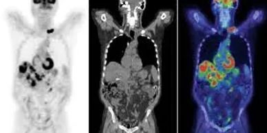
PET stands for Positron Emission Tomography. It is a procedure that produces powerful images of the human body’s biological functions. PET scans are safe and can be performed in a few hours as an outpatient procedure. Unlike conventional imaging systems such as X-rays, CTs, ultrasounds and MRIs, PET does not show body structure (anatomy). Instead, PET shows the chemical function (metabolism) of an organ or tissue. PET is used to help diagnose and treat a number of different diseases, including cancer, coronary heart disease and seizure disorders. In cancer applications, PET provides tumor imaging and has proven to be very accurate in identifying the extent of malignant disease.
CT
MRI
PET

Computed tomography, commonly known as CT or CAT scan, is a painless, non-invasive diagnostic tool that uses a specialized form of X-ray coupled with computer technology to produce cross-sectional images (slices) of soft tissue, organs, bone and blood vessels in any area of your body. CT has revolutionized medical imaging by providing more detailed information than conventional X-rays, making it possible to diagnose and manage certain diseases earlier and more accurately and, ultimately, to provide better care for patients.
MRI
MRI
X-rays

MRI is a painless, safe, noninvasive diagnostic technology that uses a strong magnet and radio waves to produce 2-D and 3-D pictures, or images, of your internal organs and other soft tissue. MRI creates images that can often provide more information than a biopsy or open surgery. MRI is also capable of creating images of biological functions, for example, the beating of your heart. Because MRI can produce clear internal images of your body from any angle, it provides doctors with a wealth of information both quickly and cost effectively. This information often cannot be obtained by any other method.
X-rays
Turn-Around Time
X-rays

X-rays are the oldest and most frequently used form of imaging to see inside the human body. X-rays are a form of radiation, like light or radio waves. They are focused into a beam that can pass through objects, including the human body. When an X-ray is done, the rays pass through the body and strike a detector, which forms an image of the inside of the body. The X-rays are absorbed by different body tissues in varying degrees. Dense tissue, like bone, absorbs most the X-rays and consequently appears white on the X-ray image. Less dense tissue absorbs less X-rays, allowing more rays to pass through and strike the detector. These tissues show on the image in various shades of gray. X-rays that pass through air, in the lungs and colon for example, are not absorbed at all. These spaces appear black on the X-ray image.
ULTRASOUND
Turn-Around Time
Turn-Around Time

Ultrasound, also called sonography, is an exam that uses high-frequency sound waves far above the range of human hearing to obtain images of the inside of the body. Sound waves are directed at a particular area of the body. The different body tissues reflect the waves back in varying degrees. The echoed waves are recorded and displayed as a continuous real-time image on a computer monitor. Since the images are real-time, ultrasound has the benefit of allowing the radiologist to see organs and tissue in motion, such as the movement of heart valves and abdominal hernias. Ultrasound relies on sound waves rather than radiation to produce images, so it is ideal in many settings. This imaging technique is becoming increasingly important in the diagnosis of medical conditions in many different organs. It is also used to evaluate pregnancy conditions.
Turn-Around Time
Turn-Around Time
Turn-Around Time
All exams are read by Anderson Radiology within 24 hours. Reports are dictated and faxed/emailed to the physician’s office. Preliminary results can be faxed to your physician’s office upon request. Reports are then placed on an FTP site for your perusal.
Copyright © 2023 Anderson Radiology - All Rights Reserved.
Powered by GoDaddy
This website uses cookies.
We use cookies to analyze website traffic and optimize your website experience. By accepting our use of cookies, your data will be aggregated with all other user data.
